Crystallography at non-ambient conditions
Crystallography at non-ambient conditions
Protein dynamics are the basis of protein function such as binding to ligands or facilitating enzymatic reactions. Motions on the femtosecond to millisecond scale determine how proteins behave ranging from bond vibrations to large scale domain movements. The corresponding free-energy landscape of proteins is defined by their chemical environment such as protein concentration, solvent, pH-value, and ligands, and also by thermodynamic parameters such as temperature, pressure, and external fields (e.g. electrical fields). Currently, protein structure determinations with X-rays and electrons are predominantly performed at cryogenic temperatures, yielding only a static picture of the frozen structures, occupying only a fraction of the conformational space accessible to the protein at physiological conditions. In contrast to electron-microscopy, structure determinations with X-rays can be also well performed at ambient conditions (room temperature or physiological temperatures, e.g. 37°C, and atmospheric pressure). However, even with these measurement opportunities, the largest part of the free-energy landscape of proteins has remained largely unexplored.
In the FS-BMX group we currently focus on studying the influence of pressure and temperature on protein structures and their dynamics.
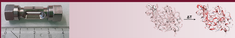
Pressure as physical variable is known to have a strong influence on the protein fold and especially on enzymatic activity. We are interested in studying the effects of pressure and temperature on protein stability and function at the atomic level with high-pressure X-ray crystallography. For this purpose, we have developed a new high-pressure cell that is pressurized with helium up to 120 MPa. With this device we are able to study several aspects that are important to understand the fundamental principles of the protein structure. We want to answer the question how proteins adapt to natural high-pressure habitats such as the deep sea, want to use pressure to better understand enzymatic mechanisms, and also aim at exploring the influence of pressure on industrially relevant enzymes. The latter will provide the fundamental basis for the rational design of a new generation of pressure-adapted enzymes, thereby fully exploring pressure as profitable process parameter for biotechnological applications.
Protein dynamics can be well studied with X-ray crystallography, as this method offers the highest spatial resolution for structure determination of macromolecules. However, these structures represent only a temporally averaged snapshot of the most populated conformations. Various other methods are available that probe protein dynamics experimentally. But they generally lack high spatial resolution. Time-resolved X-ray crystallography has begun to discern transient conformations of macromolecular structures. Here, the system is removed from its equilibrium by laser light or by addition of ligands. While the temporal resolution of ligand binding is generally hampered by the diffusion of ligands into the crystal, mostly to the millisecond range, activation by laser light can resolve femtosecond processes. Infrared laser-induced temperature jump combined with X-ray solution scattering demonstrated to be able to unveil hidden conformational dynamics in an enzyme. In contrast to solution scattering, X-ray crystallography provides a much higher spatial resolution down to the atomic level. Therefore, we combine temperature jump of protein crystals with pink beam serial crystallography to remove proteins from their conformational equilibrium. We take snapshots of the structure from nano- to milliseconds to follow the changes in the protein crystals as it returns from an energetically excited to its ground state at ambient temperatures with the aim to uncover dynamically linked networks of amino acids that could be important for protein function. This method has the potential to shed light on the protein dynamics of virtually any crystallizable protein at high spatial and temporal resolution. In the future we aim at performing such T-jump experiments in a time resolved fashion with MeV electron diffraction.
The insights gained from these projects will provide a valuable data basis for improving and extending in-silico methods for protein structure and dynamics predictions ultimately leading to a better understanding of protein function.